Week of 9/23/19
9/24
I was out sick last week, so I hadn't been able to sit down and properly inspect the plates until today.


After I took pictures of each group, I assigned each plate within the group a number (1 through 4), and noted the following:
- Number of the plate
- Who plated it
- The relative size of the colonies
- Which quadrants the colonies had spread into (in a 4-way streak)
- Colony color
- Any points of contamination
Once I had that information, I discarded the plates that had contamination and the ones that had no growth whatsoever, then decided which plates would be best used for inoculation to the next round of flasks, and which would be suitable for viewing under the dissecting microscope. Any extras were also kept, just in case.
Plates were taped together in their respective groups ("for inoculation", "for microscope", "extras") and placed in the fridge.
9/26
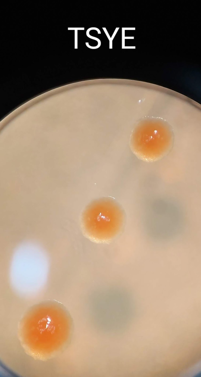
Colonies on each media were viewed under the dissecting microscope. The colonies appear to be very similar, differing primarily in size and color. However, the differences in color could be due to smaller colonies simply not having as high concentrations of bacteria to contribute to the reddish-orange color.


Comments
Post a Comment