Posts

Showing posts from December, 2019

Week of 11/25/19 - Happy Thanksgiving!

Image

Week of 11/18/19

Image
11/19 Took the PEMS inactivation plates out of the incubator. The TSB plate showed contamination and growth in quarter #2, so it will be excluded from MALDI-TOF analyzation. The other plates showed expected growth in the "live" side and no growth in the "inactivated" side. 11/21 Today I arranged the images I took from each step into documents so that I could more easily see the progression of color, clarity, and biofilm production of the flasks through the experiment. I did the same with the plate colony images to be able to contrast and compare them. 11/23 I thought back on the experiment and made notes: I would like to repeat the experiment with D. aquaticus  along with 2-3 other Deinococcus  species per discussion with Stacy. The timeline needs to be significantly tightened up. I need to make sure to ALWAYS get gram stains, particularly for bacterial growth in broth. We need to determine if there should be any changes to the media selection

Week of 11/11/19

Image
11/12 Planned what I needed to have ready in order to perform PEMS on the final batch of inoculated flasks. 11/14 Inoculated flasks from the 10/26 plates and placed them in the incubator. 11/15 Prepped the plates needed to show the inactivation of bacteria for the PEMS process. I also did some research on various Deinococcus  species. 11/16 Busy day. Flasks were taken out of the incubator and inspected for growth, then OD readings were performed. OD readings. I did gram stains for each of the flasks. The TSB sample did show contamination, indicated by the small spheres mixed in with the rods that are more typical of D. aquaticus . The gram stains also showed some discrepancies. D. aquaticus  is typically Gram negative, however, the stains I did showed a mixture of negative and positive, sometimes within the same sample. LB R2B TGY TSB TSYE With Stacy's assistance, we did PEMS on my samples to be analyzed with MALDI-T

Week of 10/28/19

Image
10/29 Much better results on this batch of plates. Only one of the LB plates showed contamination, which I attributed to excess moisture on the plates during inoculation. 10/31 I looked at the colonies found on each media under the dissecting microscope. All of them looked very similar to the first set of plates, except for TSB. The colonies were much paler in color and more spread out than before, which suggested contamination. LB R2A TGY TSB TSYE

Week of 10/21/19

Image
10/22 Melting agar and pouring plates. Lots and lots of plates. I also intended to inoculate flasks, but unfortunately, there was an issue with the autoclave and I wasn't able to get my flasks back in time. 10/24 Because of the plate contamination, I decided to essentially repeat the last two steps. I inoculated flasks from the plates from 10/5 except for those of LB and NA. I didn't have isolated colonies on those plates so I went back to the ones from 9/14. Out of curiosity, I did gram stains from the 10/5 plates, including the contamination. The contamination looks like it could be Staph. epi.  which would not be unusual. R2A TGY TSB TSYE 10/26 Flasks were removed from the incubator and inspected for growth. Unfortunately, the NA did not produce any bacterial growth, so I chose not to continue inoculations on that media since growth on NA had always been very weak. Then, I inoculated new plates from these flasks.

Week of 10/14/19

10/17 Today I planned out what media I would need to make. My flasks of TSB, TSYE, and LB broth media had to be replaced, and I needed to make more agar plates in order to repeat the inoculation that had been contaminated. 10/19 Today was all about making media. I had to make 250 mL of agar for each of the six medias, as well as 150 mL of broth for the three previously mentioned. It takes a surprising amount of time to make up that much media and of different types, so there wasn't much time for anything else.

Week of 10/7/19

Image
10/8 Took the plates we inoculated on Saturday out of the incubator to find that three of the medias were massively contaminated. All four plates for TSB, NA, and LB showed predominately white bacterial growth. The LB plates did not show any D. aquaticus growth at all. The NA plates showed very small pink colonies scattered among the white, however, none of the colonies were isolated from the white. The TSB plates showed very small coral colonies mixed with the white, with some isolated from the white. 10/10 Because of the setback of the contaminated plates, I spent most of my time planning out adjustments to my experiment and schedule.

Week of 9/30/19

Image
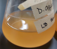
10/1 Today, Chad and I went through our box of DNA isolations and PCR products. We've been working on isolating and running PCR on each of the Deinococcus  species we have so that we can have 16s rRNA sequencing done on them. We decided that since we've had these for some time now, that Chad would run new PCR for the isolations that we already have. Then we'll only need the last two species that we currently have to be grown up, DNA isolated, and PCR run. 10/3 Today I had to make more R2B broth media, and once it was autoclaved, I inoculated from plate to flasks and placed them in the incubator. 10/4 Today, I did some planning and prep for Saturday's inoculations. John and I also mapped out an idea for a growth media database. 10/5 Today, the flasks were removed from the incubator and inspected for growth. All six showed growth, and four of the six showed biofilm formation on the sides of the flasks. Then, John an